Tendon pain can begin after one awkward landing or build gradually as training load exceeds tissue capacity. By the end, you will be able to distinguish common tendon conditions, respond safely during the first 48 hours, and recognise when symptoms require prompt specialist assessment rather than routine self-care.
Key takeaways
- A sudden snap, weakness or lost function needs prompt medical assessment.
- Rest the injured area and use ice during the first 48 hours.
- Pain that worsens with training can indicate tendinopathy from repeated loading.
- Specialist examination determines whether you need imaging, rehabilitation or further treatment.
How tendon injuries differ: tendinitis, tendinopathy, strain, tear and rupture
Pain after one forceful event suggests an acute tendon injury; pain that builds with training suggests repeated loading. A tendon strain is overstretching or microscopic damage, while a tear means some fibres have separated. A complete tear, or rupture, can cause a snap, sudden weakness and loss of function.
| Problem | What it means | Usual pattern |
|---|---|---|
| Tendinitis | Inflammation-related label for an irritated tendon | Pain and tenderness after a recent increase in activity |
| Tendinopathy | Longer-term load-related tendon change; “tendonitis” is often an imprecise name for it | Gradual pain, thickening, stiffness and pain with loading |
| Tendon strain | Overstretching or small fibre damage | One awkward movement or a sudden force |
| Partial tear | A portion of the tendon fibres has failed | Sudden pain, swelling and reduced strength, sometimes after pre-existing pain |
| Complete rupture | The tendon has structurally separated | A snap, bruising, a palpable gap or major loss of movement |
Tendinopathy often hurts during the first steps in the morning or at the start of exercise, then returns with heavier loading. Progressive pain or swelling behind the inner ankle, a flattening arch, an outward-drifting heel or inability to perform a single-leg heel raise can indicate posterior tibial tendon failure rather than a simple ankle sprain.
New tendon pain during or after a fluoroquinolone antibiotic course, especially with corticosteroid use, kidney disease or transplant history, needs prompt contact with the prescribing clinician. Avoid strenuous tendon loading until assessed.
Which sports and symptoms point to a tendon problem?
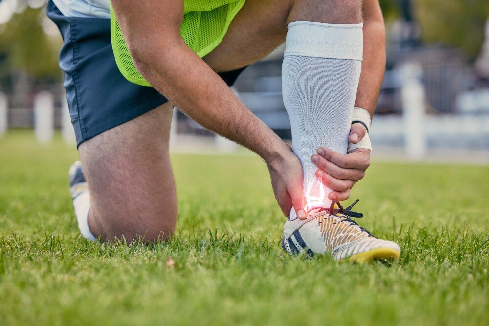
Running, jumping, racket sports and weight training all load tendons repeatedly, making them common sources of sports injuries. An overuse injury develops after repeated loading or a sudden increase in training volume; an acute injury follows one forceful landing, lift or change of direction.
| Activity | Tendon-loading pattern | Clues beyond ordinary soreness |
|---|---|---|
| Running | Repeated Achilles or patellar-tendon loading | Pain with push-off, morning stiffness, or tenderness in one precise spot |
| Jumping | Forceful landing and take-off | Swelling below the kneecap, reduced jump power, or pain that worsens with each session |
| Racket sports | Repeated gripping, serving and lunging | Localized elbow, wrist or shoulder pain that returns with the stroke |
| Weight training | Heavy, fast or poorly controlled loading | Weakness, pain during a specific lift, or a sudden snap |
| Training-volume increase | Tendons receive more work than they can recover from | Symptoms persist between sessions instead of easing after rest |
Arrange an assessment if you notice:
- Pain during loading that becomes sharper or progressively worse the next day
- Morning stiffness, visible swelling, tendon thickening or warmth
- Weakness that changes how you walk, climb stairs, grip or raise your arm
- A pop or snapping sensation followed by bruising, swelling or loss of function
- Achilles pain with inability to perform a single-leg heel raise, or inner-ankle pain with a collapsing arch
Pain during rehabilitation does not automatically mean new damage; worsening strength loss, swelling or function does.
What to do during the first 48 hours
Stop the activity that triggered the pain. First aid for tendon injury means relative rest, not complete immobility: avoid running, jumping, sprinting, heavy lifting and forceful push-off. Use a cold pack wrapped in cloth for 15–20 minutes at a time, several times during the day.
Elevate the limb and choose supportive footwear that does not bend or stretch the painful tendon.
- Do not repeatedly test strength with calf raises, hopping or forceful stretching. If the Achilles tendon may have ruptured, keep the ankle supported in a position that avoids stretching it and arrange prompt medical assessment.
- Do not massage deeply, apply aggressive stretching, or return to sport because pain has temporarily eased. These actions can enlarge a partial tear or delay recognition of a tendon rupture.
- Seek emergency care for an open wound, uncontrolled bleeding, a cold or numb foot, rapidly increasing swelling, or severe pain out of proportion to the injury.
- Contact the prescribing clinician about new tendon pain during or after a fluoroquinolone antibiotic. Avoid strenuous loading until assessed.
| Situation | Protect it now | Avoid |
|---|---|---|
| Suspected Achilles tear | Support the ankle and limit walking | Calf raises and push-off |
| Pain after an ankle sprain | Use supportive footwear and arrange assessment if pain persists behind the outer ankle | Ignoring snapping or repeated sprains |
| Pain after sudden overload | Reduce tendon loading and use a cold pack | “Pushing through” pain or high-impact exercise |
When pain needs a specialist assessment and what the examination involves
Seek emergency care for any of these signs:
- Uncontrolled bleeding, an open wound, a cold or numb foot, rapidly increasing swelling, or pain out of proportion to the injury.
Arrange prompt assessment for a suspected complete or high-grade tear, a palpable defect, major strength loss, recurrent tendon displacement, or progressive flatfoot. A sudden pop or kicked-calf sensation followed by poor push-off raises concern for an Achilles rupture; walking does not rule it out.
New tendon pain during or after a fluoroquinolone antibiotic course also needs medical review, with strenuous loading stopped until assessed.
A tendon injury specialist starts with the event, training change, medication history, pain location, and loss of function. They compare both sides, inspect swelling or deformity, feel the tendon for thickening or a gap, assess active movement and resisted strength, and watch your gait.
For suspected Achilles rupture, the Thompson test squeezes the calf while the clinician observes whether the foot points downward; an absent response supports rupture. Do not repeatedly test it with forceful stretching or calf raises.
For posterior tibial tendon dysfunction, examination includes a single-leg heel raise, arch height, heel alignment, and shoe-fit changes. Ultrasound or MRI helps when the examination cannot define the tear or when treatment planning requires detail.
In Nagpur, Dr. Girish Motwani Best Orthopedic Surgeon - SUYOG is relevant when Achilles findings or progressive flatfoot need focused foot-and-ankle assessment.
Related product
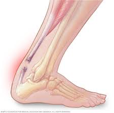 | Achilles Tendon Injuries Treatment Achilles Tendon Injuries Treatment Achilles Tendon Injuries Treatment View product → |
How diagnosis and rehabilitation guide the next step
A normal X-ray does not rule out a tendon injury: it shows fracture, avulsion, or bone abnormality better than tendon fibres. Use the test that answers the clinical question.
| Test | What it shows | When it helps |
|---|---|---|
| X-ray | Fracture, avulsion, or bone alignment | After impact, sudden pulling, or suspected bone injury |
| Ultrasound | Tendon continuity, thickening, fluid, and movement | Suspected tear or a superficial tendon injury |
| MRI | Tendon and nearby joint, muscle, and bone detail | Uncertain diagnosis, suspected partial tear, persistent symptoms, or surgical planning |
For uncomplicated tendinopathy, progressive tendon loading is usually enough. A physiotherapist may prescribe eccentric, concentric, or heavy-slow resistance exercises, then increase resistance or repetitions as strength returns. Mild exercise pain does not automatically mean further damage; reduce the load when pain worsens during exercise or the following day.
Complete rest for weeks can reduce tendon capacity.
Seek specialist care for a suspected complete or high-grade tear, palpable gap, marked weakness, open injury, recurrent tendon displacement, progressive flatfoot, or failure to improve after structured rehabilitation. Surgery is considered after the diagnosis, functional loss, and unsuccessful nonsurgical treatment are established, except for selected acute ruptures or progressive deformities.
A new pop, sudden strength loss, rapidly increasing swelling, numbness, a cold foot, or uncontrolled bleeding needs prompt or emergency assessment. Do not repeatedly stretch or perform calf raises to test a suspected Achilles rupture.
Frequently asked questions
How do tendinitis, tendinopathy, strain, tear and rupture differ?
Tendinitis describes tendon inflammation, while tendinopathy includes longer-term tendon pain and structural change. A strain involves overstretching or microscopic damage, a tear separates some fibres, and a rupture is a complete tear.
Which sports commonly cause tendon injuries?
Running, jumping, football, cricket, tennis and weight training place repeated load on tendons. A sudden forceful movement can also injure a tendon during almost any sport.
What should you do during the first 48 hours?
Stop the activity, protect the painful area, apply a wrapped ice pack for short periods, elevate the limb when practical and avoid massage or strenuous stretching. Seek urgent care for a snap, major swelling, deformity or loss of function.
When should tendon pain be assessed by a specialist?
Arrange an assessment when pain persists, worsens with activity, causes weakness or limits normal movement. Seek prompt care after a sudden snap, visible deformity or inability to bear weight or move the affected joint.
How is a tendon injury diagnosed and treated?
The examination checks tenderness, swelling, strength, movement and function. Ultrasound or MRI can clarify tendon damage when the findings are uncertain. Rehabilitation then progresses from protection and controlled movement to strengthening and a gradual return to sport.