A low arch is not automatically a medical problem: many children and adults have flexible, painless flat feet that need no treatment. Pain, stiffness, one-sided change, weakness, swelling, or declining walking ability changes the decision; by the end, you can identify those signs and understand which assessment or treatment step fits them.
Key takeaways
- Painless, flexible flat feet in young children usually need observation, not treatment.
- Arrange assessment for pain, stiffness, swelling, limping, or reduced activity.
- Assessment checks flexibility, alignment, walking pattern, strength, and footwear wear.
- Treatment may involve monitoring, exercises, orthotics, or surgery based on cause and severity.
When Is a Flat Foot a Normal Variation, and When Is It Concerning?
A painless, flexible flatfoot in a child is usually normal childhood flat feet, not a disease requiring treatment. The arch often develops during the first 5–6 years, and the foot should remain comfortable during walking, running, and play.
Check whether the arch reappears when the child stands on tiptoe or sits with the foot relaxed. That flexibility, equal appearance on both sides, and normal activity support observation; an arch that stays absent, a foot that remains stiff, or delayed motor function deserves assessment.
Arrange an assessment if the child has:
- Persistent foot or hindfoot pain
- Swelling or stiffness
- Repeated ankle sprains
- Difficulty walking, running, standing on tiptoe, or joining activities
- One foot that is flatter or more painful than the other
A rigid flatfoot is different from a flexible flatfoot. Limited side-to-side hindfoot movement, a fixed heel position, or pain with a flat foot can occur with tarsal coalition, arthritis, or another structural problem.
New or progressively worsening adult-onset flat feet deserve assessment even without dramatic pain. They can reflect progressive collapsing foot deformity, involving the posterior tibial tendon and supporting ligaments rather than arch height alone.
Weight-bearing X-rays show alignment under load when symptoms or worsening shape require investigation; uncomplicated, painless flexible flatfoot does not routinely need advanced imaging.
Which Symptoms Mean You Should Arrange an Assessment?
Arrange an assessment when flat feet hurt, stiffen, weaken, or change your walking, rather than judging concern by arch height alone. Flatfoot warning signs include persistent pain, swelling, repeated ankle sprains, reduced walking endurance, difficulty standing on tiptoe, or needing wider shoes because the foot has become broader.
| Pattern | What it may suggest | What to do |
|---|---|---|
| Pain or swelling behind the inner ankle or along the arch | Posterior tibial tendon dysfunction, ligament strain, or progressive collapsing foot deformity | Arrange a clinical assessment |
| A new or worsening one-sided flat foot | Tendon or ligament failure, arthritis, nerve weakness, or previous injury | Book an assessment, especially in adulthood |
| A rigid arch with limited subtalar movement or a heel that stays fixed | Tarsal coalition, arthritis, congenital deformity, or another structural problem | Seek specialist evaluation if painful or limiting |
| Sudden inability to bear weight, severe injury pain, rapidly increasing swelling, fever, drainage, or marked redness and warmth | Fracture, infection, or acute Charcot change | Obtain urgent medical care |
An adult flat foot that develops or progressively worsens is not simply cosmetic. A child also needs assessment for pain, asymmetry, stiffness, delayed motor skills, or difficulty keeping up with normal activities.
Do not wait for severe pain if alignment is visibly worsening or tiptoe standing has become difficult. Weight-bearing X-rays commonly clarify whether the bones and joints are collapsing under load; MRI or ultrasound is reserved for selected tendon, ligament, cartilage, or injury questions.
What Happens During a Flatfoot Assessment?
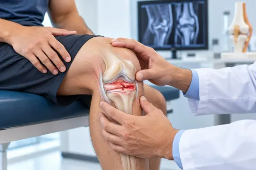
A flatfoot assessment examines how the foot functions under load, not just how high the arch looks. The clinician watches you stand and walk, checks whether the arch returns when you sit or rise onto your toes, and compares both feet for alignment, swelling, tenderness, and asymmetry.
The physical examination may include:
- A heel-rise test, usually performed on both feet and then one foot at a time. Inability to rise repeatedly on the affected side is a clue to posterior tibial tendon dysfunction, not a diagnosis by itself.
- Ankle and calf flexibility, because a tight calf or limited ankle movement can increase strain through the arch.
- Subtalar motion, heel position, muscle strength, sensation, and areas of pain.
- Whether the deformity is flexible or rigid. A fixed heel position, restricted side-to-side hindfoot movement, or an arch that stays flat while sitting or standing on tiptoe needs closer evaluation.
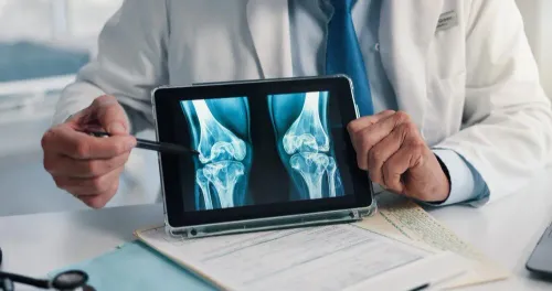
A weight-bearing foot X-ray adds useful information when pain, swelling, worsening alignment, stiffness, one-sided flattening, or reduced walking ability is present. It shows bone and joint alignment while your body is loaded, including arch collapse, heel displacement, arthritis, deformity, or a possible tarsal coalition.
Children with painful, asymmetric, or stiff flat feet may also need radiographs.
MRI or ultrasound is reserved for selected cases involving suspected tendon or ligament injury, cartilage damage, stress injury, or unexplained soft-tissue pain. Imaging is not routinely needed for an uncomplicated, painless, flexible flatfoot.
Which Treatment Step Fits the Cause and Severity?
Arch height is not the treatment target; pain, flexibility, function, cause, and progression are. A painless, flexible flatfoot in a child usually needs observation, not devices intended to create a permanent arch.
| Option | Best fit | Limit or next step |
|---|---|---|
| Observation | Painless, flexible feet with normal walking and activity | Reassess if pain, stiffness, asymmetry, or declining function appears |
| Supportive footwear | Aching feet or fatigue during standing and walking | Choose comfort and stability; shoes will not permanently raise the arch |
| Calf stretching | A tight calf limiting ankle movement or increasing strain | Use consistently; stop if it causes sharp pain |
| Physiotherapy for flat feet | Weakness, poor balance, altered gait, or reduced endurance | Follow a structured programme, not isolated exercises chosen from the internet |
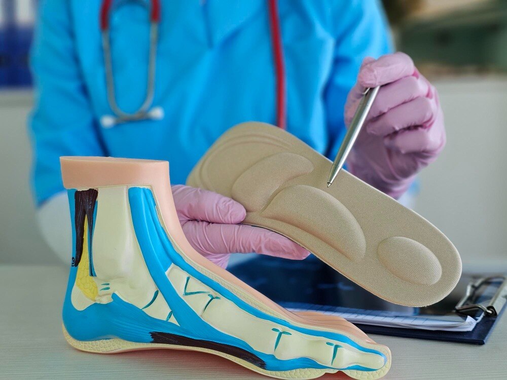
| Orthoses | Persistent symptoms despite footwear and exercises, or a clinician-identified alignment problem | Judge success by pain and function, not by a higher-looking arch |
| Surgery | Progressive deformity, a rigid or non-correctable foot, or symptoms after a reasonable nonoperative trial | Requires a cause-specific foot-and-ankle assessment |
Flatfoot treatment becomes less predictable when an adult’s alignment is worsening, the foot will not correct on tiptoe, or pain and swelling develop along the inner ankle. Repeatedly changing inserts can delay diagnosis of tendon, joint, ligament, or nerve problems.
Dr. Girish Motwani Best Orthopedic Surgeon - SUYOG can help distinguish a comfort problem from a structural or progressive condition before surgery is considered. Surgery is not justified by arch height alone, especially in a child who has no pain or activity limitation.
How Should You Monitor Flat Feet Between Assessments?
Record changes rather than arch height alone. Each week, note pain from 0 to 10, its location, swelling, stiffness, walking distance, activity limits, and whether you can rise onto each tiptoe. Photograph both feet from behind in the same position every few weeks; one-sided heel drift or increasing flattening is easier to spot this way.
Self-monitoring flat feet is reasonable when the arch has been low since childhood, both feet look similar, and you have no pain, stiffness, swelling, weakness, or loss of function. For a child, observe comfort, running, walking, and development rather than trying to build an arch with inserts.
- Stop changing inserts and arrange specialist care if pain persists or worsens.
- Seek reassessment if the foot becomes stiffer, the heel position changes, or you can no longer correct the shape by sitting or standing on tiptoe.
- Do not keep testing custom orthotics when a reasonable conservative trial has failed, adult flattening is progressing, or walking ability is declining.
Progressive collapsing foot deformity can involve the posterior tibial tendon, ligaments, Achilles tendon, and foot joints; repeated insert changes cannot identify which structure is failing. A warm, red, swollen foot with little pain is an urgent foot symptom in anyone with diabetes, neuropathy, or reduced sensation. Stop self-care and seek prompt medical assessment.
Frequently asked questions
When is a flat foot a normal variation?
A painless, flexible flat foot in a child is usually a normal variation. The arch often develops during the first five to six years, and the foot should remain comfortable during walking, running, and play.
Which symptoms mean you should arrange a flatfoot assessment?
Arrange an assessment for foot pain, stiffness, swelling, limping, repeated ankle sprains, uneven shoe wear, fatigue during activity, or reduced participation in walking, running, or play.
What happens during a flatfoot assessment?
A specialist examines foot flexibility, alignment, ankle movement, muscle strength, walking pattern, shoe wear, and symptoms. Imaging is considered when the foot is painful, stiff, asymmetric, or linked to an injury or structural problem.
Which treatments can help flat feet?
Treatment depends on the cause and severity. Options include monitoring, activity adjustments, stretching and strengthening exercises, supportive footwear, orthotics, pain relief, and surgery for selected structural or persistent cases.
How should you monitor flat feet between assessments?
Track pain, swelling, stiffness, limping, activity limits, shoe-wear changes, and whether one foot differs from the other. Arrange review if symptoms increase or daily activities become harder.