

Foot pain ranges from a minor irritation that improves with rest to a fracture, infection, circulation problem or nerve injury that needs prompt care. Use the symptom, timing and risk factors to choose between emergency assessment, same-day medical review, a routine specialist appointment and short-term home monitoring.
Key takeaways
- Seek emergency care for deformity, exposed bone, open wounds, or a cold blue foot.
- Book a specialist appointment for pain lasting weeks or returning after treatment.
- Diabetes, numbness, and poor circulation require earlier assessment of foot problems.
- Record your symptoms, injury details, footwear, medicines, and medical conditions before visiting.
Which foot-pain symptoms require emergency or same-day care?
Treat emergency foot pain as an emergency when injury causes visible deformity, exposed bone, an open wound foot injury, a cold, pale or blue foot, marked numbness, severe swelling, escalating pain or pain out of proportion to the injury.
| Level | Warning signs | Action |
|---|---|---|
| Emergency now | Cold, pale or blue foot; marked numbness; severe swelling; escalating or disproportionate pain; sudden Achilles pop with inability to push off or stand on tiptoe | Go to an emergency department |
| Emergency now | Severe pain when gently stretching the toes, especially after injury | Seek immediate care for possible compartment syndrome |
| Emergency now | New one-sided calf swelling and tenderness with chest pain or shortness of breath | Call emergency services |
| Same-day assessment | Hot, red, swollen joint with fever, chills or spreading redness; new weakness; progressive numbness; rapidly worsening pain; one-sided calf swelling and tenderness | Obtain urgent medical assessment |
| Prompt injury assessment | Inability to take four steps or inability to bear weight; tenderness at either ankle bone, the base of the fifth metatarsal or the navicular | Arrange examination and possible radiographs under the Ottawa Ankle Rules |
The Ottawa criteria still matter when you can walk: a fracture is not excluded by weight-bearing alone. A sudden pop in the back of the ankle, a kicked sensation and inability to stand on tiptoe need prompt assessment because they can indicate Achilles rupture.
Do not watch a rapidly worsening foot at home. Fever, chills, spreading redness or new neurological symptoms raise the urgency even without dramatic swelling.
When does ongoing or recurring foot pain justify a specialist appointment?
Arrange a foot-and-ankle specialist appointment when foot pain lasting more than one to two weeks continues despite reduced activity and supportive footwear. Do not wait longer if the pain repeatedly returns after walking or exercise, wakes you at night, or reduces your walking distance, work, sleep or exercise.
Mild pain still deserves review after recurrent ankle sprains, repeated ankle giving-way, persistent swelling, progressive changes in the toes or foot, or localized impact pain that keeps returning. Recurrent instability can involve torn or stretched ligaments, injured tendons or cartilage, even when each episode seems minor.
| Pain pattern | More consistent with | What to do |
|---|---|---|
| First steps after waking or rest, easing with movement | Plantar-fascia pain | Arrange review if it persists or returns |
| Pain mainly at rest or night, numbness, swelling or systemic symptoms | Atypical cause | Seek reassessment rather than repeating self-treatment |
| Focal bone pain that gradually worsens with impact | Stress fracture symptoms | Stop impact activity and arrange prompt evaluation |
A stress fracture can produce progressively sharper, localized pain while an early X-ray still looks normal. Persistent focal pain may require repeat imaging or MRI.
Night foot pain needs assessment, especially with numbness, swelling, fever or feeling unwell. These features do not fit classic plantar-fascia pain and can indicate nerve, inflammatory, infectious or bone problems.
How do diabetes, numbness and circulation problems change the threshold for care?
A diabetic foot blister, cut, callus with skin breakdown, drainage, black tissue, swelling or a change in foot shape needs prompt assessment, even without significant pain.
Diabetes, immunosuppression, a previous foot ulcer and a poor circulation foot all increase the risk of unnoticed infection, ulcer progression, fracture and Charcot-related joint collapse when protective sensation is reduced.
Do not rely only on padding, soaking or home observation. A painless wound can deepen, infect bone or destabilise the joints before you notice it.
Burning, electric or pins-and-needles pain, persistent numbness, balance changes, or toe or ankle weakness suggest nerve involvement; seek evaluation promptly when these symptoms progress or occur with back pain or radiating leg pain.
Treat these findings as urgent warning signs:
- Fever, chills, spreading redness, foul drainage or feeling unwell with a foot wound or hot, swollen foot
- Rapidly increasing swelling, black tissue, a new open area or a sudden change in foot shape
- New weakness, worsening numbness or inability to feel an injury
- A cold, pale or blue foot, or marked colour and temperature change
Before your appointment, record recent blood-glucose concerns, the wound’s onset and changes, any previous foot ulcer, and your circulation history. Note medicines that affect immunity or clotting, and take photographs if the skin changes quickly.
Which clinician should you see, and what should you record first?
Choose the care route that matches the problem, not every episode of foot pain. Use emergency services for the red flags listed earlier; otherwise:
| Route | Best fit | When to use |
|---|---|---|
| Emergency services | Serious injury or sudden deterioration | Deformity, open wound, cold or blue foot, severe numbness, escalating pain, or inability to walk |
| General practitioner for foot pain | Initial assessment | Unexplained persistent pain, possible inflammatory or systemic illness, or medication review |
| Physiotherapist foot pain | Rehabilitation | A diagnosed, clinically stable movement problem needing guided exercise |
| Podiatry service | Foot care and pressure management | Footwear problems, callus, nail or skin conditions, or pressure points |
| Foot-and-ankle orthopedic specialist | Structural or unresolved problems | Persistent or recurrent pain, instability, suspected stress injury, tendon symptoms, shape change, reduced walking tolerance, or failed rehabilitation |
Before booking, record:
- When symptoms began and the exact location
- The injury mechanism, if any, and your current walking distance
- Footwear triggers, swelling pattern, morning stiffness, numbness and fever
- Previous injuries and every current medicine
Bring the shoes you usually wear and any imaging reports. Include a written list of diabetes medicines and blood-thinning medicines. Prompt specialist review is especially important when an ankle repeatedly gives way, focal bone pain returns, or symptoms persist beyond one to two weeks despite activity modification and supportive footwear.
What happens at a foot-and-ankle specialist evaluation?
Expect an examination before imaging, not an automatic scan. A weight-bearing foot examination starts with gait and alignment while standing and walking, then focuses on the painful area. The clinician compares both sides and asks which movements, surfaces or shoes trigger symptoms.
- Observe gait and weight-bearing alignment.
- Palpate the painful area and check swelling, skin and footwear wear patterns.
- Test joint motion, tendons, strength and ankle stability.
- Perform a neurovascular foot assessment: sensation, pulses, skin temperature and colour.
Test choice follows the suspected cause. Weight-bearing X-rays, including a foot pain X-ray when appropriate, help assess fractures, alignment, arthritis and deformity. Ultrasound examines accessible tendons, fluid and some soft-tissue problems.
MRI for tendon or stress injury is more informative when an occult stress injury, tendon tear, cartilage injury or osteochondral problem is not visible on a plain radiograph. Blood tests can investigate infection or inflammatory disease.
A normal early X-ray does not settle persistent focal bone pain. The clinician may order repeat imaging or MRI because stress injuries can become clearer over time.
In Nagpur, Dr. Girish Motwani Best Orthopedic Surgeon - SUYOG can be considered when you need foot-and-ankle orthopedic assessment, examination-led testing and a plan matched to the urgency and suspected cause.
Related products
 | Ankle and foot arthritis Ankle and foot arthritis View product → |
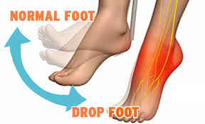 | Foot Drop Foot Drop View product → |
Frequently asked questions
Which foot-pain symptoms require emergency or same-day care?
Seek emergency or same-day care for visible deformity, exposed bone, an open wound, a cold or pale or blue foot, marked numbness, severe swelling, escalating pain, or pain out of proportion to the injury.
When does ongoing or recurring foot pain justify a specialist appointment?
Arrange a foot and ankle specialist appointment when pain persists, repeatedly returns, limits walking, follows an injury, or does not improve with sensible rest and footwear changes.
How do diabetes, numbness and circulation problems change the threshold for care?
Diabetes, reduced sensation, and circulation problems lower the threshold for assessment because a small wound or pressure area can worsen without obvious pain. Seek prompt care for skin breaks, colour changes, swelling, warmth, or drainage.
Which clinician should you see, and what should you record first?
Choose a foot and ankle orthopaedic specialist for persistent pain, injury, deformity, or suspected bone, joint, tendon, or ligament problems. Record when symptoms began, their location, triggers, severity, swelling, skin changes, footwear, treatments tried, medicines, and relevant medical conditions.
What happens at a foot-and-ankle specialist evaluation?
The evaluation includes a history, walking and alignment assessment, examination of skin, sensation, circulation, bones, joints, tendons, and range of motion. The specialist may order X-rays or other imaging when the findings require it.
Related items
 | Congenital foot anomaly like club foot,vertical talus,Hemimelia Congenital foot anomaly like club foot,vertical talus,Hemimelia Congenital foot anomaly like club foot,vertical talus,Hemimelia View product → |
 | Achilles Tendon Injuries Treatment Achilles Tendon Injuries Treatment Achilles Tendon Injuries Treatment View product → |
 | Osteochondral defects of Talus Osteochondral defects of Talus Osteochondral defects of Talus View product → |
Orthopedic Doctors Orthopaedic all orthopedic services available |